Max Mehlman is a PhD in the Psychological and Brain Sciences program.
Humans have a remarkable capacity for processing spatial information, allowing us to maintain a sense of direction and location as we navigate within our environment. This ability is largely employed unconsciously and thus often taken for granted; however, individuals suffering from chronic spatial disorientation due to brain or vestibular system damage are painfully aware of the vital contribution spatial processing makes to our quality of life. If clinicians are to better diagnose and treat these patients, we must improve our understanding of the neural mechanisms underlying spatial processing.
Recording the activity of single neurons in freely moving rodents, researchers have discovered functionally specialized cell types that encode different types of spatial information. Head direction cells fire only when the animal’s head is pointing in a specific direction within the environment – termed the preferred firing direction – and remain inactive when the animal faces outside of this direction. Individual head direction cells exhibit different preferred firing directions such that all directions are equally represented within a population of head direction cells; no matter which direction the animal is facing there will always be a subset of head direction cells that are active. Thus, the head direction cell system is continually signaling the animal’s current direction, functioning much like a compass and providing animals with a sense of direction.
Head direction cells utilize information arising independently from multiple sensory systems to accurately compute the animal’s directional heading, and by manipulating the types of information available to the animal researchers have begun to reveal how these remarkable neurons work The visual system provides head direction cells with information relating to prominent visual cues in the environment (i.e. landmarks). Head direction cells are often recorded while the animal is in a cylindrical enclosure with a single landmark attached to the inner wall. Rotating this landmark results in a corresponding shift of a head direction cell’s preferred firing direction. For example, if the landmark is moved from the north side of the cylinder to the east side, a head direction cell that initially fired when the animal faced south will now fire when the animal faces west. This finding demonstrates that head direction cells use information from the visual system to compute the animal’s directional heading relative to landmarks in the environment. However, information about landmarks – and visual information in general – is not necessary for head direction cell activity; when the landmark is removed from the environment and all other visual information is eliminated by turning off the lights, head direction cells continue to fire in a directionally-tuned manner. This is because these neurons receive inputs from the vestibular and proprioceptive systems providing information about the animal’s self-movement; this information alone suffices to support the directional firing of head direction cells.
While most rodent head direction cells reside within a well-studied set of connected brain structures called the limbic system, small numbers are found elsewhere in the brain. One such region is the dorsal striatum, which is primarily involved in motor control. Does head direction cell activity in this region depend upon output from head direction cells in the limbic system or is it generated independently? To examine this issue, I performed a brain lesion that eliminates head direction cell activity throughout the limbic system and subsequently observed the activity of neurons in the dorsal striatum. This lesion eliminated head direction cell activity in the dorsal striatum and I concluded that the activity of dorsal striatum head direction cells reflects output from the limbic system rather than functioning as an entirely independent and parallel representation of directional heading.
While my previous work demonstrated a functional relationship between the dorsal striatum and limbic system, the underlying anatomical relationship between these brain regions remained unclear. What connections in the brain permit head direction information to be conveyed from limbic system structures to the dorsal striatum?
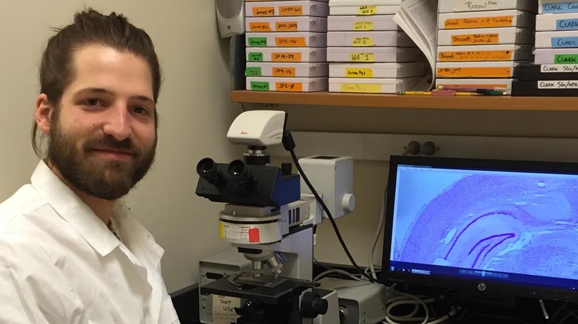
Fortunately, the Alumni Research Award allowed me to obtain the tools I needed to address this question. Using an approach called retrograde neuroanatomical tracing, I was able to visualize connections between limbic system structures and the dorsal striatum. This technique works by focally injecting a fluorescent substance into a specific brain structure; this substance – termed a tracer – is taken up only by neurons that project to the injection site. Subsequently, the brain is sectioned and a fluorescent microscope is used visualize neurons labeled with the tracer; any neurons with this label must project to the injection site. Thus, I injected a tracer into the dorsal striatum and subsequently looked for labeled neurons in the limbic system structures known to contain head direction cells. I observed robust labeling in almost every one of these structures.
This finding demonstrates that numerous head direction cell-containing structures, such as the anterior thalamus, postsubiculum and retrosplenial cortex, project directly to the dorsal striatum. These anatomical pathways represent the putative routes by which head direction information enters the dorsal striatum, ultimately driving the activity of head direction cells in this region. This finding greatly complements my previous work, and together, these studies provide the field with a more comprehensive understanding of the functional and anatomical relationships between the dorsal striatum and limbic system. Having learned this valuable neuroanatomical tracing technique, I am now using it to probe a variety of other important questions in the field.